
A person can read the smallest line on an eye chart and still have a problem with peripheral vision. That is why visual field testing explained in plain language matters: this exam checks how well you see across your full range of vision, not just directly in front of you.
At Mountain Eye Care, visual field testing is one of several diagnostic tools that can help our doctors evaluate eye health, monitor changes, and make informed recommendations. The test is straightforward, noninvasive, and often more useful than patients expect – especially when changes in vision have been gradual enough to go unnoticed.
What Is Visual Field Testing?
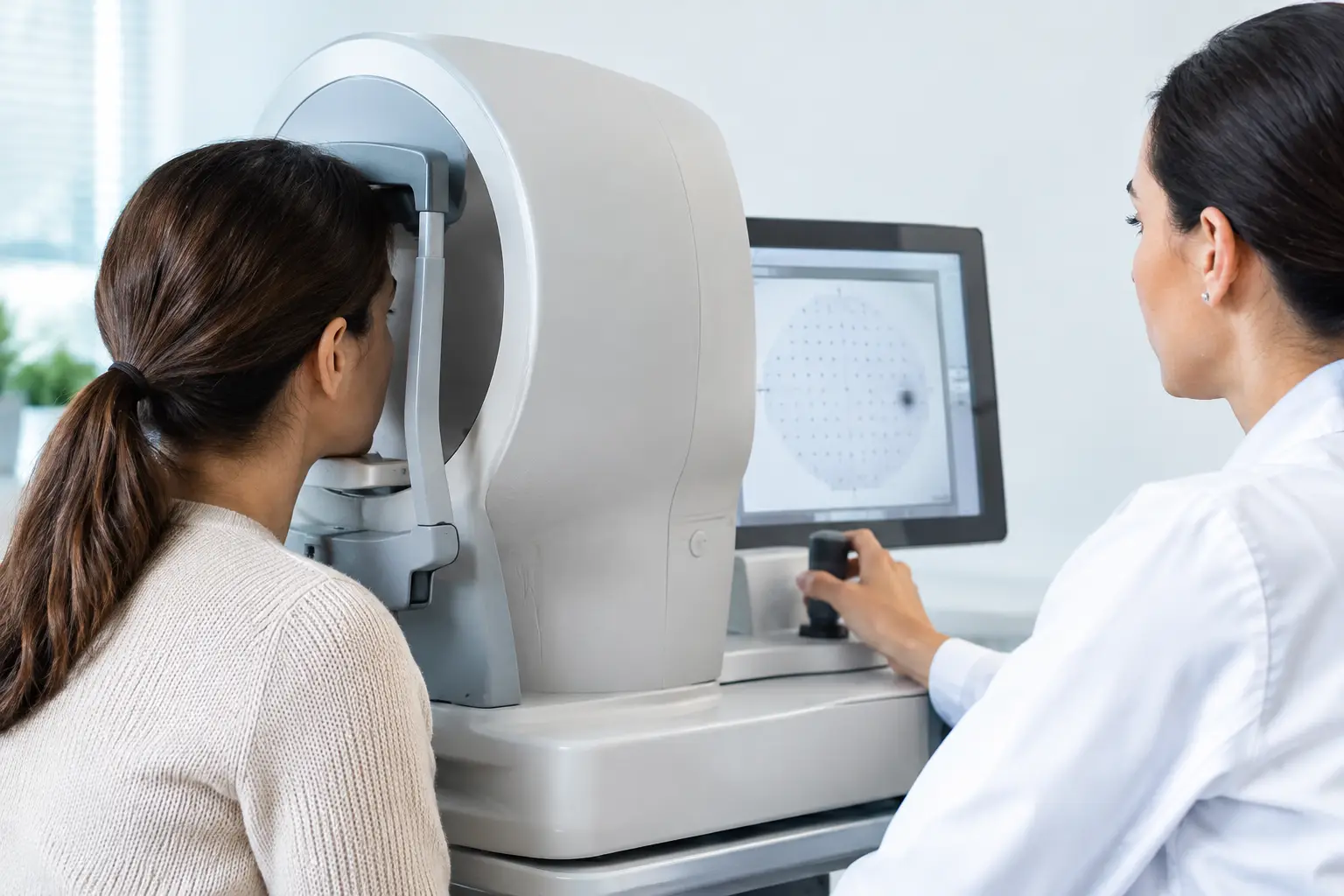
Visual field testing measures your complete field of vision: central vision, side vision, and the areas above and below your direct line of sight. During the test, you focus on a central target while small lights appear in different locations. You press a button each time you notice a light.
The computer records which lights you see and how sensitive different areas of your vision are. The result is a map of your visual field. It can show whether there are areas where vision is reduced, missing, or less sensitive than expected.
This is different from a standard visual acuity test, which measures the sharpness of your central vision. A glasses prescription may improve blur, but it cannot correct vision loss caused by damage to the optic nerve, retina, or certain parts of the brain. Visual field testing helps identify patterns that may point to those concerns.
Why Your Eye Doctor May Recommend It
Many eye conditions begin quietly. The brain is remarkably good at filling in gaps, and one eye can often compensate for changes in the other. As a result, a patient may not recognize peripheral vision loss until it becomes more advanced.
Visual field testing is commonly used to assess and monitor glaucoma. Glaucoma can damage the optic nerve, often affecting side vision first. Regular testing gives your eye doctor a baseline and allows comparison over time. A single abnormal result does not always mean glaucoma is present, but repeat testing can help determine whether a true pattern or progression exists.
The test may also be recommended when an eye examination, retinal image, OCT scan, or patient symptoms raise questions about optic nerve or retinal health. Depending on the situation, it can support evaluation of conditions such as retinal disease, optic neuritis, stroke-related vision loss, pituitary disorders, or other neurological concerns.
Symptoms that may warrant a discussion include bumping into objects, difficulty noticing cars or people from the side, missing portions of text while reading, new blind spots, or a sensation that vision is narrowing. Sudden vision loss, a new curtain-like shadow, or sudden flashes and floaters should be treated as urgent concerns rather than waiting for a routine appointment.
What Happens During the Test?
Most patients complete automated visual field testing while seated at a bowl-shaped instrument called a perimeter. One eye is tested at a time, while the other is covered. You will place your chin on a rest, keep your gaze on a central fixation light, and use a handheld button to respond whenever you see a flash of light.
The lights vary in brightness and location. Some are very easy to see; others are intentionally dim. Do not worry if you are unsure whether you saw a light. Press the button when you genuinely notice one, then return your attention to the central target.
A technician will explain the process before the test begins and can help you get positioned comfortably. Testing time varies by the type of test and the reason it was ordered, but many examinations take only a few minutes per eye. Your doctor will review the findings in the context of your complete eye exam and medical history.
You Do Not Need to “Pass” the Test
Visual field testing is not a test of alertness or a measure of effort in the usual sense. It is a clinical measurement, and it takes concentration. Blinking, fatigue, dry eyes, or learning how to respond can affect a result, particularly the first time.
Try to stay relaxed, keep looking at the center target, and avoid searching around for lights. It is normal to miss some flashes. Clicking repeatedly when no light appears can make the results less reliable, just as failing to respond to visible lights can.
If your eyes feel dry, tired, or irritated, tell the technician. A short break, a blink before starting, or rescheduling may be appropriate in some cases. Patients with dry eye symptoms may find that maintaining comfort before testing helps them focus more consistently.
How Doctors Read the Results
The printout from a visual field test can look technical, with grayscale maps, numbers, symbols, and graphs. Your doctor looks beyond any one missed spot. They assess the pattern, its severity, and whether the test itself appears reliable.
A result may show scattered missed points because a patient was tired or still getting used to the test. By contrast, a repeatable cluster of changes in a pattern consistent with optic nerve damage may need closer monitoring or treatment. Results are often compared with previous tests, which is why keeping follow-up appointments matters.
Your doctor may also compare visual field findings with intraocular pressure measurements, optic nerve appearance, retinal imaging, and OCT scans. No single test tells the whole story. This combined approach helps distinguish a temporary testing variation from a change that deserves attention.
Types of Visual Field Tests
Automated perimetry is the most common type used in comprehensive eye care. It provides detailed measurements and is particularly valuable for glaucoma monitoring. However, the best test depends on the reason for the examination.
Confrontation visual field testing is a quick screening performed by the doctor in the exam room. You may be asked to cover one eye and identify fingers or movement in different areas of your vision. This is useful for detecting larger concerns, but it is not as detailed as automated testing.
Some patients, including young children or individuals who cannot complete a computerized test reliably, may need a different approach. The goal is always to gather useful information in a way that is appropriate for the patient, not to force every patient through the same procedure.
Preparing for a More Reliable Test
There is usually little preparation required. Bring your current glasses if you use them for distance or reading, since the clinic may use a corrective lens during testing. Let the team know about any medications, recent vision changes, headaches, neurological symptoms, or prior eye diagnoses.
On the day of the appointment, arrive rested if possible. If you wear contact lenses and your eyes are uncomfortable, mention it before the test. Dilating drops can sometimes affect test timing, so your care team may perform visual field testing before dilation or schedule it separately.
The most valuable thing you can bring is patience. The test asks you to maintain focus through repetitive flashes, and that can feel demanding. A reliable test gives your doctor better information to protect your long-term vision.
When Follow-Up Testing Matters
An abnormal visual field result does not automatically mean permanent vision loss or a serious diagnosis. Technical factors, eyelid position, cataracts, and normal day-to-day variation can influence results. For this reason, doctors may repeat the test before drawing conclusions.
Follow-up becomes especially important for patients with glaucoma or those considered at higher risk because of family history, eye pressure, optic nerve appearance, or certain health conditions. The timing depends on your individual findings. Some people need periodic monitoring; others may only need testing when symptoms or exam findings change.
If visual field testing is part of your care plan, view it as a practical way to track vision that you cannot always judge at home. Showing up for recommended testing gives your eye doctor the clearest possible picture – and gives you a better chance to address changes while there is still time to act.