
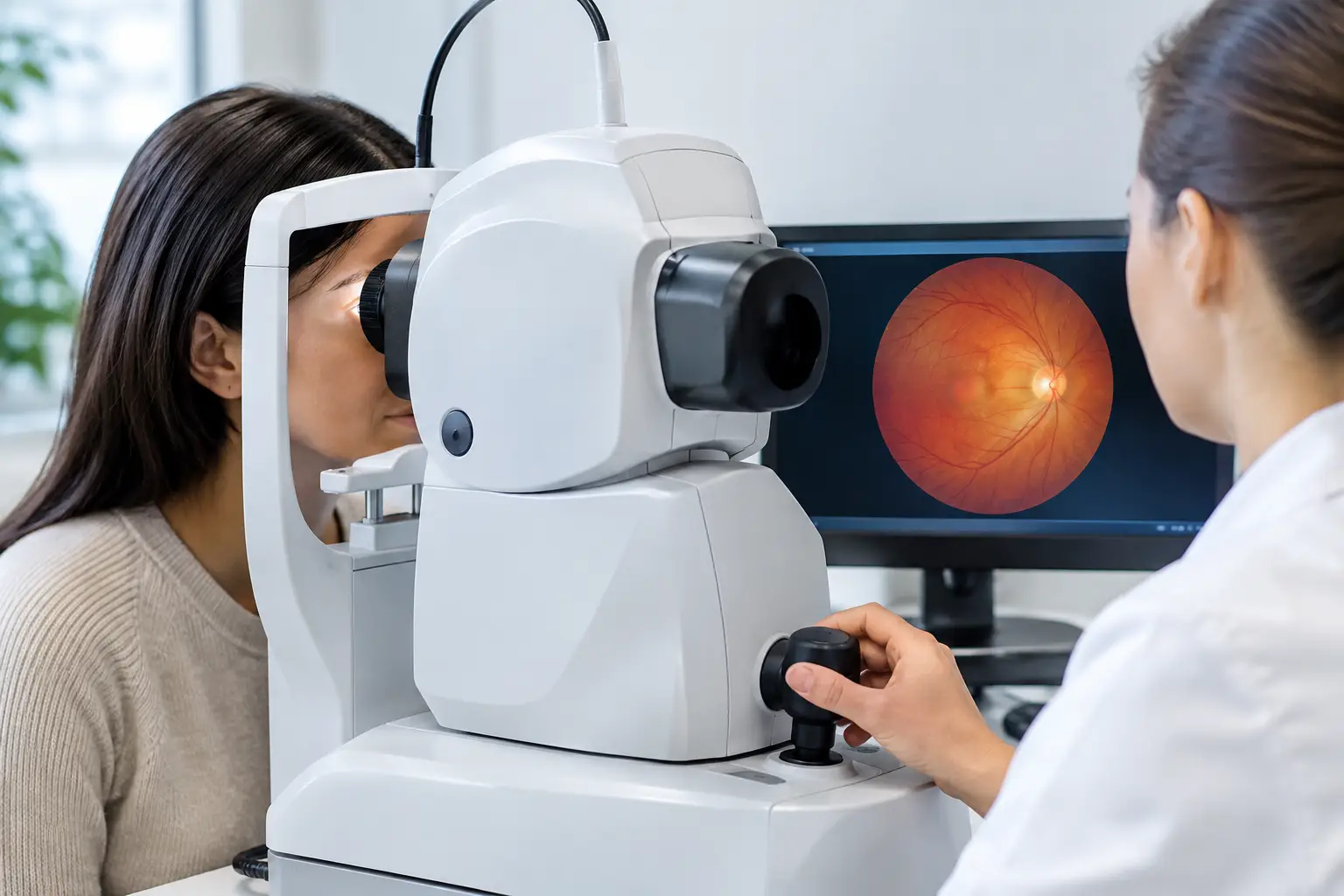
A retinal image can reveal changes in the back of the eye that you would never notice at home – often before they affect your day-to-day vision. That is why patients frequently ask what happens during retinal imaging and whether the test is uncomfortable. The short answer is that it is quick, painless, and designed to give your eye doctor a detailed view of the retina, optic nerve, and blood vessels.
For many people, retinal imaging is part of a comprehensive eye exam. It gives your doctor a useful record of your eye health today and a reliable point of comparison at future visits.
What happens during retinal imaging?
Retinal imaging uses specialized digital camera technology to photograph the inside back surface of your eye. The retina is a layer of light-sensitive tissue that sends visual information to the brain. It contains tiny blood vessels and connects closely with the optic nerve, making it an important area to assess during routine and medical eye care.
Before the image is taken, a staff member may ask you to remove your glasses. In most cases, you can keep your contact lenses in unless your doctor gives different instructions. You will sit at the imaging device, rest your chin and forehead in position, and look at a target light for a few moments.
The camera does not touch your eye. It captures one or more images while you hold still. You may notice a bright flash, similar to a camera flash, and may briefly see an afterimage afterward. That typically fades within seconds or minutes. The actual picture-taking portion usually takes only a few minutes.
Your optometrist reviews the images alongside the rest of your exam findings. This matters because an image is not interpreted in isolation. Your prescription, eye pressure, symptoms, family history, overall health, and any additional testing all help provide the full clinical picture.
Why retinal images are valuable
Many eye diseases develop quietly. Vision can feel normal while early changes are taking place in the retina or optic nerve. Retinal imaging helps doctors document and monitor findings that may be difficult to see without advanced technology.
Depending on the type of image and your individual eye health, your doctor may look for signs associated with glaucoma, diabetic eye disease, macular degeneration, retinal tears or detachments, high blood pressure, optic nerve concerns, and other retinal conditions. Imaging can also show harmless features, such as freckles or other natural variations, that are useful to record and observe over time.
The benefit is not simply getting a picture. It is being able to compare images from one appointment to the next. Small changes can be easier to identify when your doctor has a clear baseline. This is especially helpful for adults with diabetes, people with a family history of glaucoma or macular degeneration, patients who are highly nearsighted, and seniors. It can also be useful for children and adults when an eye doctor needs a more complete view of ocular health.
Retinal imaging does not guarantee that every condition will be found at its earliest possible stage, and it does not replace a comprehensive examination. Still, it is a valuable diagnostic tool that adds detail and supports more informed decisions about your care.
Will your eyes need to be dilated?
It depends on the equipment being used, the size of your pupils, and what your doctor needs to evaluate. Many modern imaging systems can capture useful retinal images without dilation. This can make the appointment more convenient, particularly for patients returning to work, school, or driving afterward.
However, dilation may still be recommended when your doctor needs a wider or clearer view of the retina. Dilating drops temporarily enlarge the pupils, allowing more light into the eye and improving visibility of areas near the edge of the retina.
If your eyes are dilated, expect blurred near vision and increased sensitivity to light for several hours. These effects vary by person. Sunglasses can make the trip home more comfortable, and some patients may prefer to arrange transportation rather than drive. Your care team can explain what is appropriate for your situation before dilation begins.
Retinal photography and OCT are not the same test
Retinal photography and optical coherence tomography, often called OCT, are both advanced eye-care tools, but they provide different information.
A retinal photograph is a color image of the surface of the back of the eye. It can document the appearance of the retina, blood vessels, optic nerve, and macula. It is particularly useful for tracking visible changes over time.
An OCT creates detailed cross-sectional images of retinal layers and, in many cases, the optic nerve. Think of it as looking beneath the surface rather than taking a standard photograph. OCT can help your doctor assess the thickness and structure of delicate eye tissues. It is often used when monitoring glaucoma, macular conditions, diabetic retinal changes, or unexplained vision concerns.
Your doctor may recommend one test, both tests, or neither test at a particular appointment. The right approach depends on your age, risk factors, symptoms, medical history, and examination findings. Advanced testing should serve a clear clinical purpose, not add unnecessary steps to your visit.
What your appointment feels like
For most patients, retinal imaging is one of the easiest parts of an eye exam. There are no needles, no recovery period, and no contact with the eye. The main challenge is simply keeping your gaze steady while the image is captured.
Young children, patients with limited mobility, and people who are sensitive to bright lights may need a little extra guidance. Let your eye care team know ahead of time if you have trouble sitting upright, focusing on lights, or keeping your eyes open because of dry eye. Small adjustments can often make the process more comfortable.
If you are having symptoms such as new flashes of light, a sudden increase in floaters, a curtain-like shadow in your vision, sudden loss of vision, or significant eye pain, do not wait for a routine imaging appointment. Seek urgent eye care promptly. Retinal imaging can be part of the evaluation, but sudden symptoms require timely professional attention.
Preparing for retinal imaging
There is usually very little you need to do in advance. Bring your current glasses and contact lens information, along with a list of medications and any relevant health updates. Be ready to share whether you have diabetes, high blood pressure, migraines, autoimmune conditions, or a family history of serious eye disease. These details can influence which tests your doctor recommends and how your images are interpreted.
If dilation may be part of your visit, consider bringing sunglasses and planning for the possibility that close-up tasks could be blurry afterward. Patients who have had dilation before can share how long the effects lasted for them, although the experience can vary from visit to visit.
At Mountain Eye Care, retinal imaging is used as part of a patient-first approach to comprehensive eye health. The goal is straightforward: provide clear information, identify concerns when they are still manageable, and create a record that supports your vision over the years ahead.
A retinal image takes only moments, but the conversation that follows can be one of the most valuable parts of your eye exam. Ask to see your images, ask what your doctor is monitoring, and make regular eye care a practical part of protecting the vision you rely on every day.