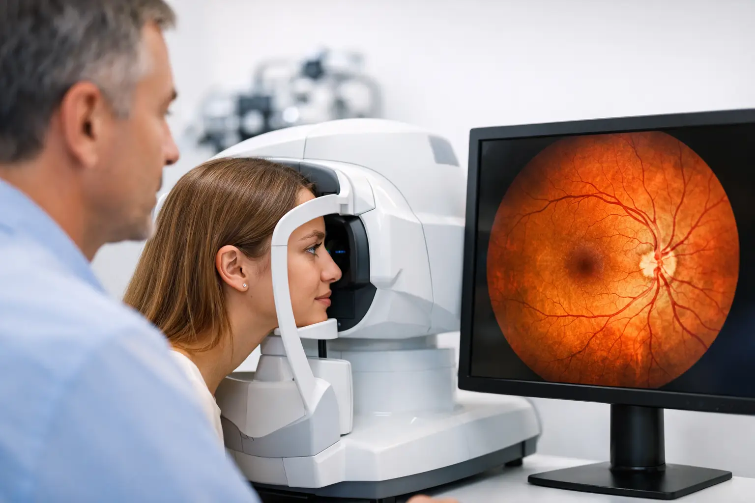
A lot can happen in the back of the eye before you notice any change in your vision. That is one reason retinal imaging for eye exams has become such a valuable part of modern eye care. It gives your optometrist a detailed view of the retina, optic nerve, and blood vessels, helping spot early signs of disease that may not cause symptoms right away.
For many patients, the technology sounds more intimidating than it actually is. In most cases, retinal imaging is quick, comfortable, and completed right in the exam room. The real benefit is not just the image itself. It is the chance to compare what your eyes look like today with what they looked like last year, which can make subtle changes easier to catch.
What retinal imaging for eye exams actually does
Retinal imaging takes high-resolution photographs or digital scans of the inside of your eye. This includes the retina, which captures light and sends visual information to the brain, as well as the optic nerve and the network of blood vessels that support eye health.
During a standard eye exam, your doctor already evaluates eye health with lenses, lights, and magnification. Retinal imaging adds another layer of detail. Instead of relying only on what is seen in the moment, your doctor can review a permanent record of the back of the eye, enlarge specific areas, and monitor changes over time.
That matters because many eye conditions develop gradually. Early diabetic eye disease, glaucoma-related optic nerve changes, macular degeneration, retinal tears, and even signs of high blood pressure can sometimes be visible before a patient notices blurriness, pain, or vision loss.
Why doctors recommend it
The strongest case for retinal imaging is early detection. Eye disease does not always announce itself with obvious symptoms. Some of the most serious conditions progress quietly, and by the time vision changes appear, damage may already be harder to treat.
Retinal imaging can help identify concerns sooner and support better decisions about follow-up testing or treatment. It can also reduce uncertainty. If an eye looks healthy, the image becomes part of your baseline record. If something appears suspicious, your doctor has a clearer way to explain what they are seeing and why monitoring matters.
For families, this can be especially helpful. Parents often want reassurance during a child’s exam, while older adults may be monitoring age-related changes or health conditions such as diabetes. A visual record gives everyone a more concrete picture of eye health.
What happens during the test
In most offices, retinal imaging is simple. You sit at a machine and look at a target while the camera captures images. The test usually takes just a few moments per eye.
Some systems can take images without dilation, while others work better if the pupils are widened with eye drops. Whether dilation is needed depends on the equipment, your pupil size, and how much of the retina your doctor needs to examine. If dilation is recommended, it is not because something is wrong. It may simply provide a better view.
The image process itself is painless. You might notice a bright flash of light, similar to a camera. That is often the most uncomfortable part, and even then, it passes quickly.
Retinal imaging versus dilation
Patients often ask whether retinal imaging replaces dilation. Sometimes the answer is yes for a routine screening image, but not always.
Retinal imaging is an excellent tool, but it does not eliminate the need for a doctor’s clinical judgment. Dilation gives a wider, more direct view of the retina, especially the outer edges where tears or other peripheral problems can occur. If you have symptoms such as flashes, floaters, sudden vision changes, or risk factors for retinal disease, dilation may still be the better choice, or your doctor may recommend both.
This is where the trade-off matters. Imaging is convenient and creates a lasting record. Dilation can provide a broader examination of structures that may be harder to capture in a single image. The best option depends on your age, symptoms, medical history, and the reason for the visit.
Conditions retinal imaging may help detect
A retinal image does not diagnose every problem by itself, but it can reveal signs that need attention. That includes diabetic retinopathy, where blood vessels leak or swell; glaucoma, where the optic nerve may show subtle damage; and macular degeneration, which affects central vision.
It may also reveal retinal hemorrhages, pigment changes, suspicious lesions, swelling, or signs of systemic health issues. In some cases, eye findings lead patients to follow up with their primary care physician because the retina can reflect changes linked to diabetes, hypertension, and vascular disease.
That is one reason advanced eye exams are about more than updating a glasses prescription. Your eyes can provide important clues about your overall health.
Who benefits most from retinal imaging for eye exams
Retinal imaging can be useful for almost anyone, but it tends to be especially valuable for patients at higher risk. That includes people with diabetes, high blood pressure, a family history of glaucoma, past retinal problems, strong prescriptions, or age-related eye disease concerns.
Children and teens can benefit too, especially when doctors want a baseline record or a closer look without making the exam more stressful. Adults who prefer to avoid dilation for work or scheduling reasons often appreciate non-dilated imaging when it is appropriate.
It is also helpful for patients who are monitoring a known issue over time. Comparing this year’s image with earlier scans can show whether a spot, vessel pattern, or optic nerve appearance has remained stable or changed enough to warrant further testing.
How this fits into a comprehensive eye exam
Retinal imaging works best as part of a full exam, not as a stand-alone shortcut. A comprehensive visit still includes vision testing, refraction, eye pressure measurement when indicated, and a doctor’s evaluation of both the front and back of the eye.
In some cases, your doctor may recommend additional technology such as OCT, which provides cross-sectional images of retinal layers, or visual field testing, which checks for functional vision loss. Each test serves a different purpose. Retinal imaging offers a broad photographic record. OCT gives a more detailed structural view beneath the surface. One is not automatically better than the other. It depends on what your doctor needs to evaluate.
Practices that invest in this level of diagnostic technology are usually focused on catching problems early and managing care more precisely. At Mountain Eye Care, that patient-first approach supports both routine exams and more complex eye health needs.
Common questions patients have
One common concern is cost. Some practices include retinal imaging in certain exam packages, while others offer it as an optional add-on. The value often comes down to your risk factors and how useful a baseline image will be in future care. If you are unsure, ask how the test may change your exam or support long-term monitoring.
Another question is whether the test is safe. For the vast majority of patients, yes. It is noninvasive and uses light to capture the image. If a bright flash bothers you, the discomfort is brief.
Patients also ask whether a normal image means everything is fine. A healthy retinal photo is reassuring, but no single test catches every issue. That is why regular exams still matter, especially if you have symptoms or ongoing medical concerns.
When it is worth saying yes
If your doctor recommends retinal imaging, there is usually a good reason. Maybe they want a baseline. Maybe you have a health condition that raises your risk. Maybe your eyes are healthy today, and they want the clearest record possible to help keep them that way.
The bigger point is simple. Eye exams are no longer limited to reading letters across the room. With better imaging technology, doctors can see more, explain more, and track more over time. For patients, that means fewer guesses and a better chance of protecting vision before problems become harder to ignore.
If you have been putting off an exam because your vision seems fine, this is a good reminder that eye health and eyesight are not always the same thing. Sometimes the most valuable part of the visit is finding out what you cannot feel yet.