
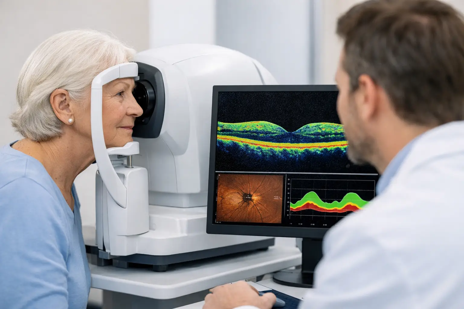
Glaucoma often develops quietly. Many people can have early optic nerve damage without pain, without blurred vision, and without any obvious warning signs during daily life. That is exactly why an oct scan for glaucoma detection has become such an important part of modern eye care. It gives your eye doctor a detailed look at structures inside the eye that can show signs of damage before you notice a problem yourself.
For patients, that matters because glaucoma is not just about eye pressure. Pressure is one piece of the picture, but it does not tell the whole story. Some people have elevated pressure and never develop meaningful damage. Others have pressure in a normal range and still lose nerve tissue over time. The goal is not simply to find a number that looks high. The goal is to protect vision by identifying change early and following it carefully.
What an OCT scan for glaucoma detection actually does
OCT stands for optical coherence tomography. The scan uses light waves to create high-resolution cross-sectional images of the retina and optic nerve. In glaucoma care, doctors use OCT to measure the thickness of the retinal nerve fiber layer and the ganglion cell layer, both of which can become thinner when glaucoma damages the eye.
The test itself is quick and noninvasive. You sit at the instrument, look at a target, and the device captures images in seconds. There are no injections, no contact with the eye in most cases, and no recovery time afterward. For many patients, it feels much easier than they expected.
What makes OCT especially useful is that it turns structures we cannot judge precisely with the naked eye into measurable data. Your doctor is not just looking for a vague impression that the nerve seems different. The scan provides thickness maps, symmetry comparisons, and progression analysis that support earlier and more confident decisions.
Why glaucoma can be missed without detailed imaging
A standard eye exam can reveal many signs of glaucoma risk, including elevated eye pressure, thinning of the optic nerve rim, or changes on visual field testing. Even so, early glaucoma can be subtle. Structural damage may begin before a patient notices any blind spots, and sometimes before visual field testing shows a clear defect.
That gap is where OCT becomes valuable. It can detect thinning in vulnerable tissue before vision loss becomes obvious in day-to-day life. It does not replace a full glaucoma workup, but it strengthens it. When doctors combine OCT with pressure readings, optic nerve evaluation, corneal thickness measurements, and visual field testing, they get a much clearer understanding of whether glaucoma is present, suspected, or progressing.
This is also why one normal pressure reading does not automatically mean everything is fine. Glaucoma is a disease of optic nerve damage, not just a disease of high pressure. A patient may have borderline findings in one area and more convincing findings in another. Careful diagnosis depends on seeing the whole picture.
What your doctor looks for on an OCT scan
During an OCT review, one major focus is the retinal nerve fiber layer around the optic nerve. In many glaucoma cases, this layer becomes thinner in specific patterns. The machine compares your measurements to an age-based reference database, which helps identify whether parts of the nerve tissue fall outside an expected range.
Doctors also look at the ganglion cell complex in the macula. This can be particularly helpful when damage is early or when the optic nerve appearance is difficult to interpret. In some patients, ganglion cell loss may support suspicion even when other findings are not yet dramatic.
Equally important is whether the scan is reliable. A result is only useful if the image quality is good and the scan was centered properly. Dry eye, cataracts, poor fixation, small pupils, or simple movement during the test can affect the data. That is one reason glaucoma diagnosis should never depend on a printout alone. Experienced interpretation matters.
An OCT scan for glaucoma detection is powerful, but not a stand-alone test
Patients sometimes hear that OCT can find glaucoma early and assume it gives a simple yes-or-no answer. In practice, it is more nuanced than that. OCT is one of the best tools we have for structural assessment, but it does not diagnose every case by itself.
There are several reasons. Some healthy eyes naturally vary from the reference database. Highly nearsighted eyes can produce measurements that look suspicious even when glaucoma is not present. A tilted optic nerve or certain retinal conditions can also make interpretation more complicated. On the other side, very early damage may still be difficult to confirm on a single scan.
That is why follow-up matters. In glaucoma care, change over time is often just as important as one isolated result. If repeated OCT scans show progressive thinning, that can be a strong sign that the optic nerve is under stress. If scans remain stable and other testing is normal, the concern may be lower. This long-term view is a major part of protecting sight.
Who should consider OCT testing for glaucoma
Not every patient needs the same level of testing at every visit, but OCT is especially useful for people with higher risk. That includes patients with elevated eye pressure, a family history of glaucoma, suspicious optic nerve appearance, thin corneas, diabetes, or a history of visual field changes. It may also be recommended more often for older adults, since glaucoma risk rises with age.
Some patients are surprised to learn that family history carries real weight. If a parent or sibling has glaucoma, your own risk is higher. That does not mean you will definitely develop it, but it does mean screening should be taken seriously. Glaucoma-related vision loss is permanent, so waiting for symptoms is not a safe strategy.
For patients already diagnosed with glaucoma, OCT helps establish a baseline and monitor progression. Treatment decisions are often based on whether the disease appears stable. If pressure looks acceptable but the OCT suggests continued nerve loss, the treatment plan may need to change.
What to expect during the appointment
If your doctor recommends OCT as part of a glaucoma evaluation, the process is straightforward. A technician may first review your history, measure your vision, and check eye pressure. Depending on the exam, your pupils may or may not need to be dilated. Then the OCT scan is performed at the imaging device.
Afterward, the doctor reviews the images in context with the rest of the exam. This is the part patients should value most. The benefit is not just taking the scan. The benefit is having the results interpreted alongside optic nerve appearance, corneal thickness, symptoms, medication history, and any visual field findings.
In a well-equipped practice, this makes the visit more efficient and more informative. Instead of relying on guesswork or waiting for obvious loss, your doctor can make decisions using objective structural data.
Why early detection changes the long-term outlook
Glaucoma damage cannot be reversed, but it can often be slowed significantly when caught early. That is the practical value of OCT. It helps move care from reacting to advanced vision loss to identifying subtle change before daily function is affected.
For patients, that can mean preserving driving vision, reading comfort, mobility, and independence for years longer. For doctors, it means more confidence in deciding when to observe, when to repeat testing, and when to begin or adjust treatment.
This is particularly reassuring for patients who have been told they are glaucoma suspects. That label does not always mean disease is present, but it does mean closer attention is warranted. With modern imaging and regular monitoring, a suspicious finding does not have to become a missed opportunity.
At Mountain Eye Care, advanced imaging is part of a broader commitment to careful, patient-first eye health management. When technology is paired with experienced clinical judgment, patients get clearer answers and more personalized follow-up.
If you have a family history of glaucoma, rising eye pressure, or simply have not had a comprehensive eye exam in a while, asking whether OCT testing is appropriate is a smart next step. Sometimes the most important part of protecting your vision is finding what you cannot feel yet.